
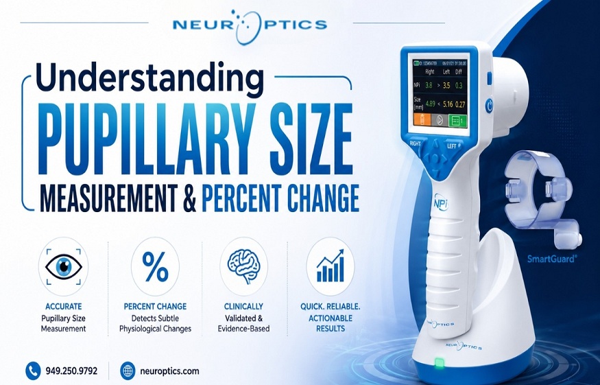
Understanding Pupillary Size Measurement& Percent Change
What are thehospitals really missing in pupillary assessment?
Walk into any neurology unit, and you’ll find nurses doing what they’ve always done – shining a pen light into a patient’s eyes, estimating size, noting “reactive” or “sluggish,” and moving on. It works, until it doesn’t.
The problem isn’t clinical skill. It’s that manual pupillary evaluation was never built for the precision modern neurocritical care demands. A 0.5 mm change, a subtle asymmetry, a reactivity shift at 2 a.m. – these details get lost in estimation. And in neurology, lost details cost patients.
Subjective Assessment and Its Clinical Consequences
Pupil response reflects brainstem activity, cranial nerve integrity, and intracranial pressure in real time. That makes it one of the most clinically loaded assessments a bedside nurse performs; yet it’s still largely subjective.
Two trained clinicians examining the same patient routinely document different sizes. Neither is wrong. Both are estimating. That variability creates documentation gaps, inconsistent escalation, and missed windows for early intervention.
Several conditions directly influence what clinicians observe during pupillary sizemeasurement:
- Traumatic or ischemic brain injury– often producing fixed or asymmetric dilation.
- Glaucoma– where sustained intraocular pressure changes affect reactivity over time.
- Opioids, sedatives, or atropine– each altering size and response in distinct ways.
- Anxiety and sympathetic activation– causing transient bilateral dilation that can mimic pathology.
Getting this right means knowing not just what the pupil looks like now, but how it’s changed, and how fast.
Dilated Pupils and Stroke: What Hospitals Need to Know
A unilateral fixed dilated pupil in a declining patient is textbook uncal herniation. It can also indicate an eye stroke or hemorrhagic expansion. Bilateral dilation reads differently – metabolic causes, pharmacological effects, or diffuse cerebral dysfunction are all possibilities.
Neither presentation can be fully interpreted from size alone. Reactivity, symmetry, and the percent change in pupil size over a defined period tell a far more complete story. That’s the clinical data manual assessment that simply can’t be generated consistently.
How NPi Scoring Strengthens Clinical Decision-Making
The NPi scores pupillary reactivity from 0 to 5 using constriction velocity, dilation velocity, latency, and amplitude. It’s not a replacement for clinical judgment; it’s the objective layer underneath it.
In a structured neuro exam, a drop from 3.5 to 2.8 is quantifiable, documentable, and actionable, even when nothing looks visually different to the eye. Research consistently links NPi scores below 3 to elevated intracranial pressure in TBI and post-cardiac arrest populations. That’s a specific, measurable threshold nurses can act on, and physicians can trust.
For hospital operations, the downstream value is real: fewer escalation delays, tighter documentation, defensible records, and better-equipped nursing staff.
A Clinical Scenario worth Examining
Post-operative neurosurgical patient. Aneurysm clipping. Manual checks every four hours, both pupils are reactive and stable at around 3 mm. At hour two, automated pupillometry records an NPi of 2.6 on the left, down from 3.4. The percent change in pupil size has shifted from baseline.
Read More: How Dietitians Play an Important Role in Helping People Stay Healthy?
The CT scan confirms early vasospasm. Treatment is initiated promptly, well before any neurological deficit has the chance to develop, and the patient recovers fully.
That’s not luck. That’s what objective neurological tools are built to catch.
Frequently Asked Questions
Q: Is it normal for pupils to change size?
A:Mild fluctuation with light or medication is normal. Unilateral or sustained change paired with neurological signs is not.
Q: Can anxiety change pupil size?
A:It can. Sympathetic activation dilates pupils temporarily. In monitored patients, objective data separates this from a true neurological event.
Q: What eye conditions affect pupil size?
A:Horner’s syndrome, Adie’s tonic pupil, third nerve palsy, and various drug effects all alter pupillary response. Consistent pupillary size measurement is what makes differentiation possible.
Q: When should hospitals be concerned about dilated pupils?
A:When dilation is unilateral, fixed, or accompanied by altered consciousness or asymmetry, especially in post-surgical or head trauma patients.
Closing Thoughts
Manual assessment isn’t going away. But relying on it alone in high-acuity neurological care is a gap hospitals can no longer afford to overlook. Objective pupillometry gives clinical teams the data to act earlier, document better, and make decisions with confidence, not estimation.
The tools exist. The evidence is there. The next step is simply deciding to use them.
Ready to Standardize Pupillary Assessment at Your Hospital?
Explore how NeurOptics pupillometry solutions bring objective, real-time pupillary evaluation to neurocritical care teams, and help hospitals catch what the pen light misses.
 Understanding Pupillary Size Measurement& Percent Change
  Why Natural-Looking Enhancements Are Changing the Conversation Around Beauty
  Treatment for Depression and Addiction: Why Both Need Care at the Same Time
  How Dietitians Play an Important Role in Helping People Stay Healthy?